Centrocyte vs Centroblast: Reading Germinal Center Cytology
1 October 2026 · By Dr. Maitrayee Roy and Dr. Akshay Bali
Three Cells, One Follicle
Every germinal center on your slide has these three players at work, and the exam loves asking you to tell them apart.
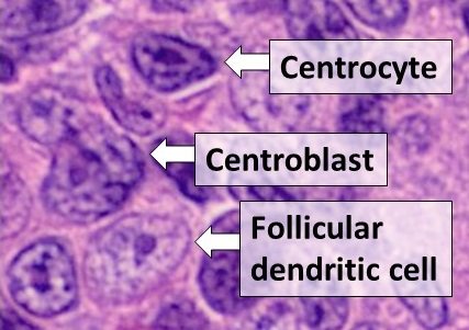
Centroblasts sit in the dark zone — large cells with open, vesicular nuclei, several peripheral nucleoli, and scant basophilic cytoplasm. They're the proliferating population: high mitotic rate, actively rearranging and hypermutating their immunoglobulin genes.
Centrocytes sit in the light zone — smaller, with irregular, cleaved or angulated nuclei and denser chromatin. These are the cells undergoing selection: if a centrocyte's newly mutated antibody doesn't bind antigen well enough, it's meant to die by apoptosis. If it does bind well, it survives and moves on to become a memory B cell or plasma cell.
Follicular dendritic cells (FDCs) aren't B cells at all — they're stromal cells that form a meshwork through the germinal center, trapping antigen-antibody complexes on their surface (CD21+, CD23+, CD35+) and presenting that antigen to centrocytes during selection. On H&E they're easy to miss; they have pale, oval nuclei with delicate chromatin and inconspicuous cytoplasm, blending into the background unless you're specifically looking for the meshwork pattern.
Why This Distinction Actually Matters on the Exam
Here's the pearl that turns this from a labeling exercise into a diagnosis: in a normal, reactive germinal center, these B cells are BCL-2 negative. The selection process depends on apoptosis actually working — cells that fail selection need to die, and BCL-2 (an anti-apoptotic protein) would block that.
In follicular lymphoma, the t(14;18) translocation puts BCL2 under the control of the immunoglobulin heavy chain promoter, so the neoplastic germinal center B cells switch BCL-2 on. That single IHC stain — BCL-2 positive within follicles — is one of the fastest ways to flag a follicular proliferation as neoplastic rather than reactive, especially when combined with loss of the normal dark zone/light zone polarization and back-to-back, poorly-defined follicles replacing the normal nodal architecture.
See follicles on low power → check architecture and polarization → if anything looks off, BCL-2 is your next stain, not your last resort.
Quick Self-Check
- Large cell, open nucleus, peripheral nucleoli, scant cytoplasm → centroblast
- Smaller cell, cleaved/irregular nucleus, denser chromatin → centrocyte
- Pale oval nucleus, forms a meshwork, not a true lymphocyte → follicular dendritic cell
- BCL-2 positive within follicles → think follicular lymphoma, not reactive hyperplasia
FAQ
Q: Is BCL-2 positivity alone enough to diagnose follicular lymphoma?
A: No — it's a strong supporting finding, but diagnosis integrates architecture (loss of polarization, back-to-back follicles, effacement of normal nodal structure), cytology, and often BCL6/CD10 co-expression and molecular confirmation of the t(14;18) translocation where needed.
Q: Why are follicular dendritic cells relevant if they're not even lymphocytes?
A: Because CD21/CD23 IHC for FDCs shows a disrupted, expanded, or irregular meshwork in follicular lymphoma versus the tight, well-organized meshwork of a normal reactive follicle — it's a second architectural clue beyond BCL-2 alone.
Go Deeper
This exact distinction shows up across FRCPath Part 1, FRCPath Part 2, and NEET SS Pathology — it's one of the panels in our free pathology comic book (“BCL-2 isn’t on that report for decoration — it’s the actual reason the cell refuses to die on schedule.”)
It's covered in full in the hematopathology module of the FRCPath Part 1 course
Practice this exact kind of reasoning with the 1200+ MCQ bank